

Knee Cartilage
Demo Author One
Cartilage repair options for lasting joint health
A practical overview of cartilage repair, injection therapy, rehabilitation planning, and the clinical evidence that guides joint preservation care.
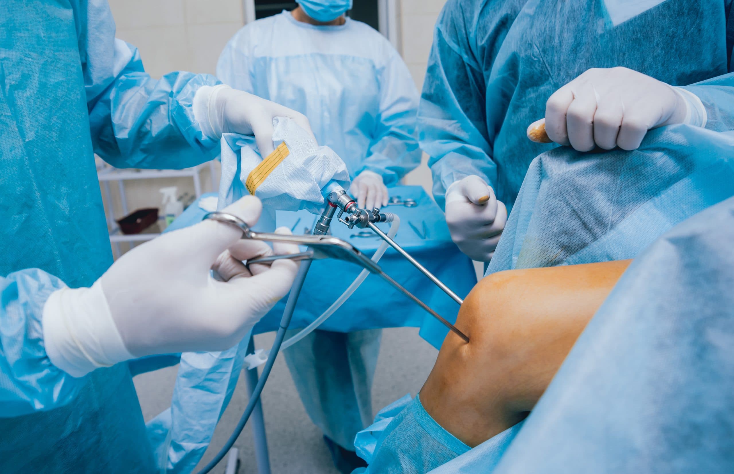
The evidence
A plain-English look at the evidence behind STACi — hyaline-like cartilage formation, MRI outcomes and safety.
 Reviewed byProf Paul Lee MBBch, FRCS (Tr & Orth), PhDLast reviewed 1 May 2026
STACi evidence comes from the class of single-stage, scaffold-based cartilage cell therapies (including the INSTRUCT-generation trials). Studies of this approach report lasting improvement in pain and function, formation of durable hyaline-like cartilage in most patients, good defect fill on MRI, and a safety profile comparable to other cartilage treatments. Individual results depend on the defect.
STACi belongs to a well-studied family of treatments: autologous chondrocyte therapies, in which your own cartilage cells (chondrocytes, KON-droh-sites) are used to regrow a joint surface. The original two-stage techniques, ACi and MACi, have been studied for more than two decades. STACi is the single-stage, three-dimensional-scaffold evolution of that lineage — the laboratory step is brought into theatre so cells are harvested, processed and implanted in one operation.
Because STACi shares its biology with these established therapies, the sensible way to read its evidence is in two layers: the large, long-running body of work on autologous chondrocyte implantation as a whole, and the newer single-stage, scaffold-based (INSTRUCT-class) studies that test the specific approach STACi uses. Both point the same way — a living cartilage repair, not a temporary patch.
Professor Lee frames every STACi around five things the new tissue needs to take hold and last — the reason the procedure is assessed and tailored rather than sold as one-size.
Your own cartilage cells — the living material that becomes new cartilage.
The 3D scaffold the cells grow through, in depth as well as across the surface.
Your own biological boosters (PRF, mFat or BMAC where indicated) that feed the cells.
A stable, well-prepared joint — right alignment, clean defect rim, no ongoing instability.
A staged rehabilitation programme that loads the joint at the right pace to mature the tissue.
The most important question about any cartilage repair is what actually grows back. Microfracture, the simplest option, tends to fill a defect with fibrocartilage — a weaker, scar-type tissue that does not wear as well as the original. The point of cell-based regeneration is to do better: to form hyaline-like cartilage, the smooth, load-bearing surface a healthy joint is built from.
Evidence for scaffold-based autologous chondrocyte therapy reports formation of hyaline-like cartilage in the majority of treated defects, with the three-dimensional scaffold supporting growth through the full depth of the damage rather than only skinning the surface. Where biopsies and MRI have been examined, the repair tissue is described as maturing and integrating with the surrounding cartilage over the first year — consistent with the staged recovery STACi patients follow.
Cartilage repair is tracked on MRI using a standardised score called MOCART (Magnetic resonance Observation of Cartilage Repair Tissue), which grades how well a repair has filled the defect, how smoothly its surface sits, and how completely it has bonded to the surrounding cartilage and bone. It is the standard way researchers compare cartilage procedures objectively rather than on symptoms alone.
Single-stage scaffold cartilage therapies of STACi’s class report good defect fill and integration on MOCART assessment, improving over the first year as the tissue matures.
Pain, function and return to activity
STACi is a regeneration procedure, not a quick painkiller. The measured goal is durable improvement in pain and everyday function, tracked with validated knee scores such as the IKDC (International Knee Documentation Committee) score and the KOOS (Knee injury and Osteoarthritis Outcome Score). Studies of single-stage, scaffold-based cartilage cell therapy report meaningful, lasting gains on these measures, with many patients returning to their previous activity level.
Your individual result depends on the size, depth and location of the damage, the health of the surrounding cartilage, and how closely rehabilitation is followed. We tell you, in writing at consultation, what to expect for your specific defect — and every outcome feeds our ICRS-aligned cartilage regeneration database.
Because STACi uses your own cells and your own biological augmentation, it avoids the rejection and disease-transmission concerns of donor material. The evidence for autologous chondrocyte therapies describes a safety profile comparable to, or better than, other cartilage treatments, with the main considerations being those common to any joint surgery — swelling, stiffness and the need to protect the graft while it beds in. Delivering STACi as a single operation also removes the second surgery and second anaesthetic that two-stage ACi and MACi require.

STACi belongs to a well-studied family of autologous chondrocyte therapies, and the single-stage, scaffold-based (INSTRUCT-class) approach it uses has been tested in its own right. That evidence reports lasting improvement in pain and function and the formation of durable, hyaline-like cartilage in most patients. Your imaging review at consultation sets realistic expectations for your specific defect.
The aim, supported by the evidence, is hyaline-like cartilage — the smooth, load-bearing surface a healthy joint is built from — rather than the weaker fibrocartilage that microfracture tends to produce. The 3D scaffold supports growth through the full depth of the defect.
MOCART is a standardised MRI score that grades how well a cartilage repair has filled the defect, how smooth its surface is, and how completely it has integrated. It lets clinicians compare cartilage procedures objectively rather than on symptoms alone.
Cartilage repair is judged over years, not weeks, because the tissue matures over the first year and durability is what matters. We give you written, defect-specific expectations at consultation and record every outcome in our ICRS-aligned database.
It uses your own cells and biological augmentation, avoiding rejection and donor-tissue risks. The evidence describes a safety profile comparable to or better than other cartilage treatments, with the usual considerations of any joint surgery. Single-stage delivery removes a second operation and anaesthetic.
Still have more specific concerns?
Free Discovery CallThe only way to know what STACi could achieve for you is an imaging review. Talk it through first on a free discovery call, or book a consultation with the surgeon who will perform the procedure.
London Cartilage Clinic
Clinical updates, cartilage treatment guidance, and recovery-focused articles from our specialist team.
A practical overview of cartilage repair, injection therapy, rehabilitation planning, and the clinical evidence that guides joint preservation care.
A practical overview of cartilage repair, injection therapy, rehabilitation planning, and the clinical evidence that guides joint preservation care.
A practical overview of cartilage repair, injection therapy, rehabilitation planning, and the clinical evidence that guides joint preservation care.